Researchers from the University of Basel have identified a novel deep muscle layer within the masseter—the key elevator muscle of the lower jaw essential for chewing—that has never been formally described before.
The masseter is the primary muscle for mastication. It elevates the mandible against the maxilla to generate chewing force, contributes to mandibular protrusion and lateral movements, and subtly influences facial aesthetics.
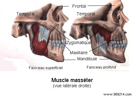
Standard modern anatomy texts describe the masseter as having two layers: superficial and deep. However, historical accounts have hinted at a possible third layer, though descriptions of its location remain inconsistent.
In a rigorous anatomical study, University of Basel researchers dissected formaldehyde-preserved human heads from a dozen specimens, conducted CT scans on sixteen cadavers, and analyzed an MRI from a living subject to confirm this elusive third layer's presence and morphology. Their findings appear in the journal Annals of Anatomy.
The study conclusively isolated an anatomically distinct deep third layer of the masseter, extending from the medial surface of the zygomatic process of the temporal bone to the root and posterior border of the coronoid process on the mandible.
"This deep portion of the masseter is distinctly separate from the other two layers in both trajectory and function," explains lead author Szilvia Mezey. "The fiber arrangement suggests it stabilizes the mandible by elevating and retracting the coronoid process." Notably, this layer is the only component of the masseter capable of pulling the jaw backward.
"While anatomy research over the last century was thought exhaustive, this finding echoes the thrill of zoologists discovering a new vertebrate species," adds co-author Jens Christoph Türp.

To standardize references, the researchers propose naming it M. masseter pars coronoidea (coronoid portion of the masseter). This discovery could refine jaw surgeries and treatments for temporomandibular joint disorders.