Harvard researchers used advanced fluorescent imaging to create detailed maps of microbial communities on the human tongue.
Bacteria are ubiquitous—on door handles, keyboards, phones, and even within our bodies. Beyond skin microbes, the majority thrive in the digestive tract, including the esophagus, stomach, colon, and mouth. Today, we focus on the warm, moist environment of the oral cavity.
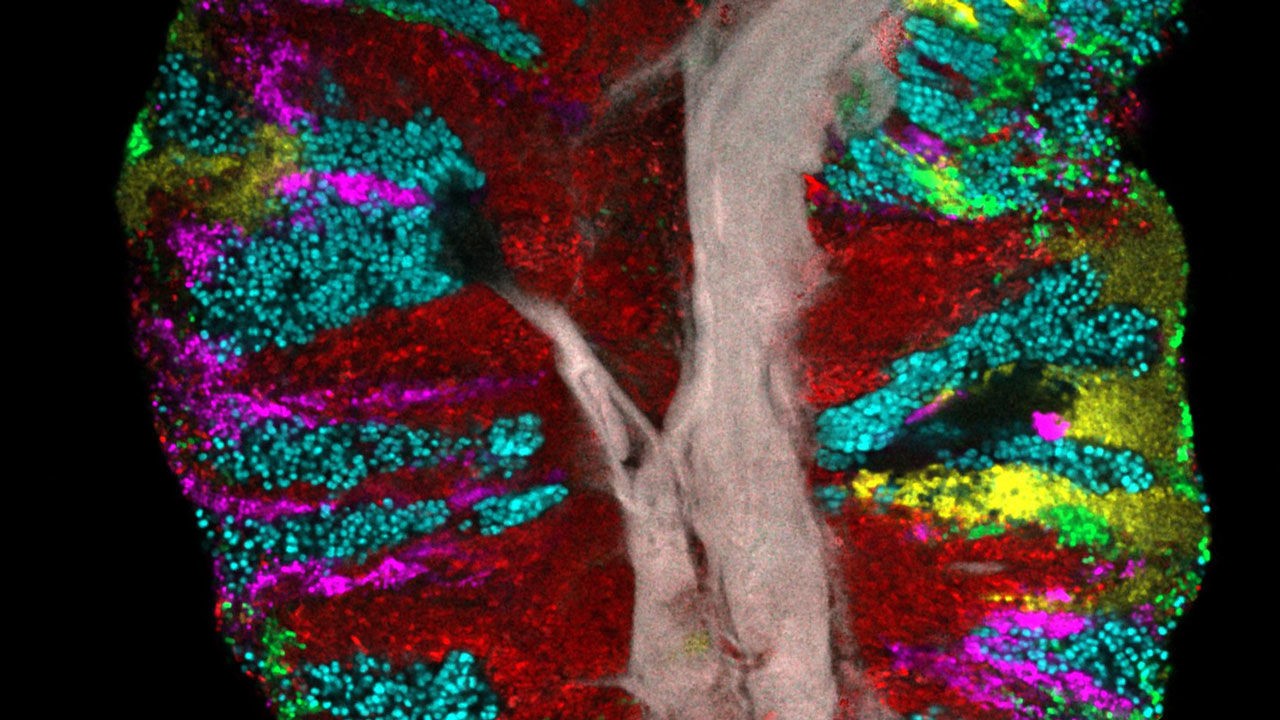
While various bacterial species inhabit the mouth, their spatial organization remained elusive—until now. Scientists at Harvard School of Dental Medicine developed an innovative fluorescent imaging technique to analyze tongue samples from 21 human subjects, isolating distinct microbial communities.
They identified 17 bacterial genera present in over 80% of individuals. Notably, bacteria form species-specific clusters, as shown in the image below highlighting the three most abundant groups. Actinomyces (red) clusters near the tongue's epithelial tissue (gray), Rothia (sky blue) forms large aggregates between communities, and streptococci (green) gather along the edges.
These maps enable researchers to track how microbial colonies form and evolve, shedding light on their roles in oral health and beyond.
Just as the gut microbiome influences mood and mental health, disruptions in the oral microbiome may contribute to systemic conditions. A June 2019 Science Advances study found that mice infected with P. gingivalis—a gum disease pathogen—developed Alzheimer's-linked brain proteins.
Earlier research linked tongue microbiome changes to pancreatic cancer, suggesting oral microbiota monitoring could enable earlier detection.
This underscores why what enters our mouths matters profoundly, making such studies invaluable.
Source