A remarkable scanning electron microscope image from a U.S. laboratory captures SARS-CoV-2 virus particles budding from a dying host cell.
Once SARS-CoV-2 enters the body, its viral particles target respiratory tract cells. They fuse their lipid membranes with the host cell's, injecting genetic material inside.
The cell then produces numerous viral copies. Once replicated sufficiently, these clones burst out from the dying cell to infect new hosts.
In essence, SARS-CoV-2 hijacks cells, turning them into virus factories before discarding them as they die.
This striking image, taken by Elizabeth Fischer, head of electron microscopy at Rocky Mountain Laboratories in Montana, depicts "viral shedding"—the moment virus particles emerge from an infected cell.
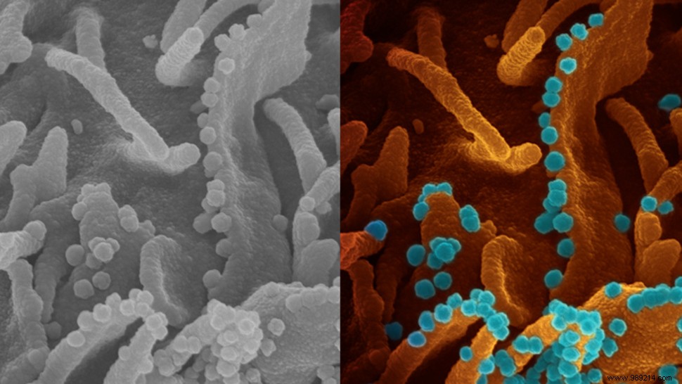
Fischer explains: "The folds and orange-brown protrusions are part of the surface of a single cell infected with SARS-CoV-2. This cell is from a commonly studied primate renal epithelial cell line. The small blue spheres emerging from the cell surface are SARS-CoV-2 particles."
The image highlights SARS-CoV-2's remarkable replication efficiency: a single infected cell can release thousands of new particles, each ready to infect others.
Viral shedding from the infected host enables transmission to others. For SARS-CoV-2, the primary route is the respiratory tract via coughs and sneezes.
COVID-19's basic reproduction number (R0) is estimated at 2.2 to 3.9, meaning one infected person typically spreads it to about three others—fueling rapid outbreaks. This viral affinity for human cell receptors, combined with efficient shedding, explains its swift spread.
By comparison, influenza's R0 ranges from 0.9 to 2.1, while measles is 12 to 18, underscoring its extreme contagiousness.
Source