Medical professionals rely on advanced imaging techniques to examine the body internally. The most common methods—ultrasound, X-ray, CT, PET, and MRI—provide detailed insights for accurate diagnosis.
Ultrasound (Echography)
Ultrasound is frequently used for abdominal issues and prenatal care. High-frequency sound waves reflect off body tissues, creating real-time video images visible on a screen.
X-ray
X-rays are the go-to for detecting bone fractures. Radiation passes through the body, producing shadow-like images of bones, organs, and tissues. They're also valuable for assessing heart failure, pneumonia, or lung tumors.
CT (Computed Tomography)
Building on X-ray technology, CT scans capture multiple cross-sectional images, which computers reconstruct into detailed 3D views. This allows thorough evaluation of organs, bones, and soft tissues.
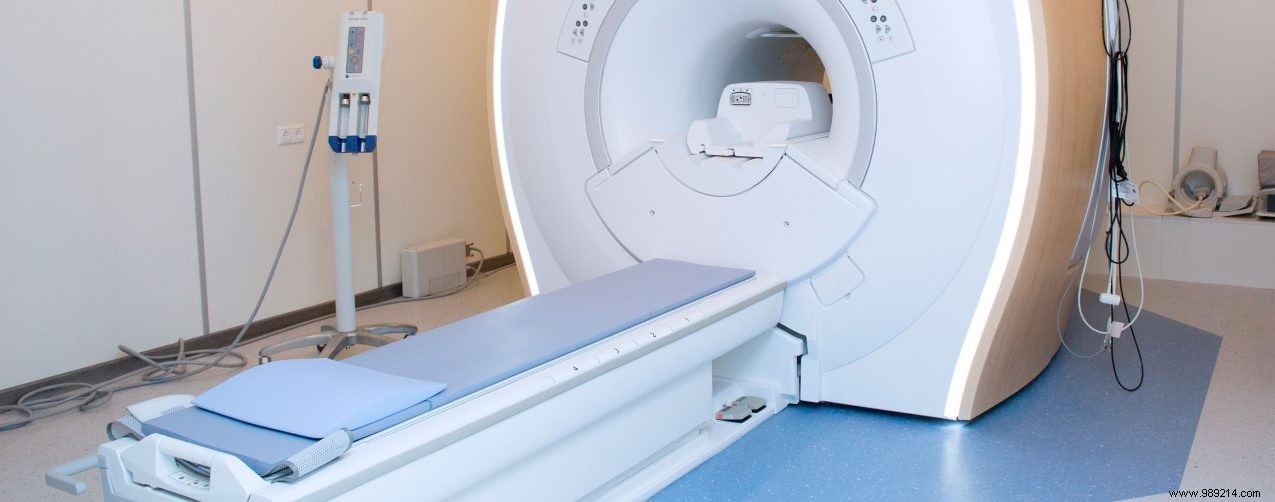
MRI (Magnetic Resonance Imaging)
MRI avoids radiation, using powerful magnets and radio waves instead. The magnet aligns the body's hydrogen atoms, and varying tissue compositions produce distinct signals for slice-by-slice computer-generated images. It's especially effective for brain and neurological assessments.
PET (Positron Emission Tomography)
A small amount of radioactive tracer is injected, which spreads through the body and emits detectable radiation. PET excels at identifying infections, inflammation, and metabolic activity, often outperforming other methods per recent studies.